CORNEAL AND OCULAR SURFACE PROCEDURES-Pterygium excision with autograft with fibrin glue- Single Eye cost in Iran
CORNEAL AND OCULAR SURFACE PROCEDURES-Pterygium excision with autograft with fibrin glue:
A pterygium is a tissue growth in the corner of the eye that is often triangular in shape. If the growth is not addressed, it can spread over the pupil, obstructing vision or distorting the surface of the eye, resulting in hazy vision.
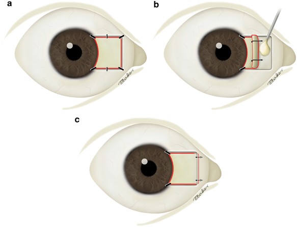
Pterygium surgery involves removing noncancerous conjunctival growths (pterygia) from the eye. The clear tissue that covers the white area of the eye and the inside of the eyelids is known as the conjunctiva. A pterygium might cause little to no symptoms in some circumstances.
The autograft includes transplanting a small piece of the patient's conjunctiva into the location where the pterygium will be removed. To avoid the necessity for sutures, absorbable sutures or tissue adhesive (biological glue) are utilised.
Fibrin glue (also known as fibrin sealant) is a surgical preparation that is used to generate a fibrin clot for hemostasis or wound healing. Human fibrinogen and human thrombin are packaged individually.
Disease Overview:
Noncancerous conjunctiva growths (pterygia)
On the cornea, a pterygium is a benign development of fibrovascular tissue. These lesions can grow red and inflammatory, encroach on the visual axis, and cause sufferers aesthetic and functional problems. Pterygia most commonly affects people between the ages of 20 and 50.
Disease Signs and Symptoms:
The first signs of a pterygium are redness and thickness in the corner of the eye, generally the one closest to the nose. The growth can spread over the eye's surface and into the iris (the coloured part of the eye). A person may detect the development of a pterygium but show no other signs or symptoms. Other symptoms that may be noticed include:
- Redness and inflammation of the eyes
- An abrasive or burning sensation in the eyes
- A sensation as if there were something strange in your eye
- Reduced tear production causes eye dryness.
- If the translucent layer at the front of the eye (cornea) is deformed, vision blurs.
- If growth encroaches over the pupil, vision will be obscured.
Disease Causes:
The most common cause of pterygia is excessive exposure to ultraviolet (UV) radiation. This is more frequent in those who live in sunny places or who work in jobs that expose them to UV rays (eg: farmers, fishermen, arc welders).
Disease Diagnosis:
The diagnosis of a pterygium is simple. A physical examination with a slit light may be used by your eye doctor to identify this issue. With the use of magnification and strong illumination, this lamp helps your doctor to examine your eye.
Disease Treatment:
A pterygium does not always create difficulties or necessitate treatment. If the pterygium is causing discomfort or obstructing vision, there are two basic treatment options to consider.
Medication
Topical corticosteroid eye drops can be used to relieve redness and irritation for a short period of time. Artificial tears are used to keep the eye adequately moisturised when dryness is an issue.
If your vision is damaged or your symptoms are especially bothersome, surgery may be considered.
The pterygium is carefully removed during surgery, and a portion of conjunctiva from beneath the eyelid is transplanted into the location where the pterygium was. The surgery is done under local anaesthesia and takes around 30 minutes to complete.
Pterygia can return following surgery, however this happens in a tiny number of instances.
Country wise cost comparison for CORNEAL AND OCULAR SURFACE PROCEDURES-Pterygium excision with autograft with fibrin glue- Single Eye:
| Country | Cost |
|---|---|
| India | $587 |
| Iran | $1436 |
Treatment and Cost
3
Total Days
In Country
- 1 Day in Hospital
- 2 No. Travelers
- 2 Days Outside Hospital
Treatment cost starts from
$1474
Popular Hospital & Clinic
Featured Hospital
0 Hospitals
Related Packages
CORNEAL AND OCULAR SURFACE PROCEDURES-Pterygium excision with autograft with fibrin glue- Single Eye
Start from in India
$587 $618
5% off
CORNEAL AND OCULAR SURFACE PROCEDURES-Pterygium excision with autograft with fibrin glue- Single Eye
Start from in Iran
$1436 $1474
5% off