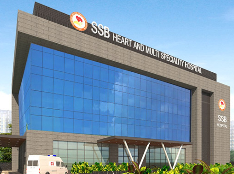
SSB Heart and Multispecialty Hospital, Haryana, India
Balloon Pulmonary Valvuloplasty
SSB Heart and Multispecialty Hospital, Haryana, India
-
Our Price USD 2428
-
Hospital Price USD 2556
-
You Save : USD 128
Booking Amount: USD 243. Pay Remaining 90% at the hospital.
- Explore benefits & save additional 30%
Additional Credit
Among the important extras we offer as part of the Additional Credit are the following:
-
Site Tourism For The Patient & Attendant
-
Airport Pick & Drop Service
-
Ambulance service at airport
-
Priority appointments with The Doctor
-
Cancel Easily Anytime with Full Refund
-
Room Upgradation
-
Free Online Doctor Consultation Valued at USD 20
-
Free hotel Stay for 5 to 7 days Accordingly
-
Welcome Kit at Arrival
-
Interpreter
-
Medical Visa Assistance
What is Included?
- Doctor consultation charges
- Lab tests and diagnostic charges
- Room charges inside hospital during the procedure
- Surgeon Fee
- Cost of implant
- Nursing charges
- Hospital surgery suite charges
- Anesthesia charges
- Routine medicines and routine consumables (bandages, dressings etc.)
- Food and Beverages inside hospital stay for patient and one attendant.
What is not Included?
- Extra Radiology Investigations
- Healthcare Professionals Charges of other consultations.
- Other Requested Services such as Laundry etc.
- Additional Pharmaceutical Products and Medicines After Discharge from Hospital.
- Management of Conditions Unrelated to Procedures or Pre-Existing.
- The cost of any additional implants will be in addition to the package cost.
Package Description
Balloon Pulmonary Valvuloplasty
Balloon valvuloplasty is a procedure that tries to repair the pulmonary valve. A catheter is a long, thin tube that is used in the process. At the end of this tube is an inflated balloon. This catheter is threaded into a blood artery in the groyne and all the way to the pulmonary valve by the healthcare professional. After then, the balloon is inflated. It helps open the valve by stretching it. Blood may then flow freely to the pulmonary artery without being obstructed.
This technique is designed to alleviate the symptoms of congenital pulmonary stenosis. Valvuloplasty isn't necessary for many persons with this problem. Mild instances may not show any signs or symptoms. When exercising, however, if you have a mild case, you may experience fatigue and shortness of breath.It's possible that you won't experience any symptoms at first. However, you may develop them later in life, either as a youngster or as an adult. Severe symptoms frequently necessitate treatment with a procedure or surgery. Pregnant women are frequently advised to seek treatment, especially if the stenosis is severe.
If you do require surgery, your doctor is most likely to prescribe balloon valvuloplasty. It's a far less intrusive procedure than open-heart surgery. Recovery time is frequently shortened as well.
Disease Overview
Pulmonary valve stenosis
A narrowing of the valve between the lower right heart chamber (right ventricle) and the lung arteries is known as pulmonary valve stenosis (pulmonary arteries). The valve flaps (cusps) of a constricted heart valve might become thick or rigid. Blood flow via the valve is reduced as a result of this.
Usually, pulmonary valve disease develops as a result of a cardiac condition that occurs prior to birth (congenital heart defect). Adults, on the other hand, may acquire pulmonary valve stenosis as a side effect of another condition.
The severity of pulmonary valve stenosis varies. Some persons with moderate pulmonary valve stenosis have no symptoms and just need to see their doctor once in a while. Moderate and severe pulmonary valve stenosis may necessitate a valve repair or replacement operation.
Disease Signs and Symptoms
The indications and symptoms of pulmonary valve stenosis vary depending on how much blood flow is obstructed. Mild pulmonary stenosis causes no symptoms in some persons. Those with severe pulmonary stenosis may experience symptoms for the first time when exercising.
Symptoms and indicators of pulmonary valve stenosis include:
- murmur, a whooshing sound that may be heard using a stethoscope.
- Fatigue
- Breathlessness, especially with physical exercise
- Pain in the chest
- Consciousness loss (fainting)
Blue babies can have pulmonary valve stenosis and other congenital heart abnormalities (cyanotic).
Disease Causes
The most common cause of pulmonary valve stenosis is a congenital cardiac abnormality. The actual reason for this is unknown. As the kid grows in the pregnancy, the pulmonary valve does not develop properly.
The pulmonary valve is made up of three flaps, which are thin bits of tissue (cusps). With each pulse, the cusps open and close, ensuring that blood flows in the appropriate direction.
One or more cusps may be stiff or thick in pulmonary valve stenosis, or the cusps may be linked (fused) together. The valve does not fully open as a result of this. The reduc
The right ventricle's pressure rises as it tries to force blood through the narrower aperture. The increasing pressure puts a strain on the heart, causing the muscular wall of the right ventricle to thicken.ed valve opening makes blood flow out of the lower right heart chamber more difficult (right ventricle).
The following conditions or illnesses may raise the likelihood of pulmonary valve stenosis:
- German measles (rubella)
- Noonan syndrome
- Rheumatic fever
- Carcinoid syndrome
Diagnosis
Children are frequently diagnosed with pulmonary valve stenosis. However, it is possible that it will not be recognised until later in life.
The doctor will listen to your or your child's heart with a stethoscope. It's possible to hear a whooshing sound (murmur) created by choppy (turbulent) blood flow past the constricted valve.
The following tests may be used to identify pulmonary valve stenosis:
Echocardiogram: An echocardiography is a type of ultrasonography of the heart. Sound waves are employed to produce moving images of the heart. An echocardiography allows doctors to examine the aortic valve and aorta in greater detail. It can assist in determining the source of aortic valve disease as well as the severity of the condition.
Electrocardiogram (ECG or EKG): The electrical activity of the heart is recorded in this noninvasive examination.
Cardiac catheterization: This procedure rarely used to diagnose aortic valve dysfunction. It can, however, be used to assess the degree of aortic valve disease. This can provide further information regarding blood flow and how effectively the heart is functioning.
Other Imaging tests: To confirm the diagnosis of pulmonary valve stenosis, magnetic resonance imaging (MRI) and computed tomography (CT) scans are sometimes
Disease Treatment
If you have minor pulmonary valve stenosis and don't have any symptoms, you may just need to see your doctor once in a while.
You may require a cardiac treatment or heart surgery if you have moderate or severe pulmonary valve stenosis. Your general health and the look of your pulmonary valve will determine which operation or surgery you will undergo.
The following heart treatments and surgeries are used to treat pulmonary valve stenosis:
Balloon valvuloplasty: A flexible tube (catheter) with a balloon on the tip is inserted into an artery, generally in the groyne, by the doctor. X-rays are utilised to guide the catheter to the heart's restricted valve. The doctor inflates the balloon to enlarge the valve opening and, if necessary, separate the valve flaps. After that, the balloon is deflated. The catheter and balloon are withdrawn from the patient.
Valvuloplasty can help to increase blood flow through the heart and relieve the symptoms of pulmonary valve stenosis. The valve may, however, narrow again. In the future, some people will require valve maintenance or replacement.
Pulmonary valve replacement: If balloon valvuloplasty isn't a possibility, the pulmonary valve may be replaced by open-heart surgery or a catheter technique. If there are any additional congenital cardiac problems, the doctor can usually correct them all at the same time.
Antibiotics are required before some dental operations or surgeries for those who have had pulmonary valve replacement to avoid endocarditis.
utilized.
Information related to Treatment
Package Details
Days in Hospital
3 Days
Days in Hotel
*
10 Days
Room Type
Private