VITREO RETINAL PROCEDURES Pars Plana Viterctomy 25G for Single Eye, India
VITREO RETINAL PROCEDURES Pars Plana Viterctomy 25G for Single Eye
India
-
Our Price USD 2095
-
Hospital Price USD 2205
-
You Save : USD 110
Booking Amount: USD 209. Pay Remaining 90% at the hospital.
Book NowAdditional Credit
Among the important extras we offer as part of the Additional Credit are the following:
-
Site Tourism For The Patient & Attendant
-
Airport Pick & Drop Service
-
Ambulance service at airport
-
Priority appointments with The Doctor
-
Cancel Easily Anytime with Full Refund
-
Room Upgradation
-
Free Online Doctor Consultation Valued at USD 20
-
Free hotel Stay for 5 to 7 days Accordingly
-
Welcome Kit at Arrival
-
Interpreter
-
Medical Visa Assistance
What is Included?
- Doctor consultation charges
- Lab tests and diagnostic charges
- Room charges inside hospital during the procedure
- Surgeon Fee
- Cost of implant
- Nursing charges
- Hospital surgery suite charges
- Anesthesia charges
- Routine medicines and routine consumables (bandages, dressings etc.)
- Food and Beverages inside hospital stay for patient and one attendant.
What is not Included?
- Extra Radiology Investigations
- Healthcare Professionals Charges of other consultations.
- Other Requested Services such as Laundry etc.
- Additional Pharmaceutical Products and Medicines After Discharge from Hospital.
- Management of Conditions Unrelated to Procedures or Pre-Existing.
- The cost of any additional implants will be in addition to the package cost.
Package Description
VITREO RETINAL PROCEDURES Pars Plana Viterctomy 25G:
Retina surgeons employ pars plana vitrectomy (PPV) to execute a number of surgeries. The treatment begins with the removal of the "vitreous gel" that fills the back of the eye (hence the term "vitrectomy").
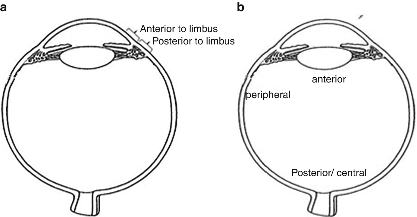
The pars plana, a "safe zone" in the white area of the eye or sclera, is typically used to enter the dilated eye; hence, the technique is known as a pars plana vitrectomy. A surgical microscope with a specific lens provides a wide view as well as a magnified and detailed image of the interior of the eye.
The pars plana vitrectomy (PPV) is a vitreoretinal surgical procedure that allows access to the posterior segment in a controlled, closed system to treat problems such retinal detachments, vitreous haemorrhage, endophthalmitis, and macular holes.
For many weeks after surgery, your eye may be puffy, red, or irritated. For a few days following the procedure, you may experience eye discomfort and your eyesight may be impaired. Before you can resume your usual activities, you will need to heal for 2 to 4 weeks.
Disease Overview:
Retinal detachment is a condition in which a thin layer of tissue at the back of the eye (the retina) slips away from its usual position. Retinal detachment occurs when the layer of blood vessels that supplies oxygen and sustenance to the retina separates from the retinal cells.
The longer you wait to treat a retinal detachment, the more likely you are to lose vision in the afflicted eye permanently.
The abrupt emergence of floaters and flashes, as well as impaired vision, are all possible symptoms of retinal detachment. Contacting an ophthalmologist (eye doctor) as soon as possible will help you save your vision.
Disease Signs and Symptoms:
The process of retinal detachment is painless. However, there are virtually always warning signals before it develops or has progressed, such as:
- The unexpected emergence of a large number of floaters – little specks that appear to wander across your field of vision
- Light flashes in one or both eyes (photopsia)
- Vision is hazy
- Reduced side (peripheral) vision with time
- Over your visual field, there is a curtain-like shadow.
Disease Causes:
Retinal detachment may be classified into three types:
Rhegmatogenous: These are the most prevalent forms of retinal detachments. A hole or tear in the retina allows fluid to travel through and gather behind the retina, forcing the retina away from the underlying tissues, causing rhegmatogenous detachments. The portions of the retina that detach lose blood flow and stop operating, resulting in visual loss.
Tractional. When scar tissue forms on the surface of the retina, the retina pulls away from the back of the eye, producing detachment. People with poorly managed diabetes or other diseases are more likely to develop tractional separation.
Exudative. Fluid collects behind the retina in this form of detachment, but there are no holes or tears in the retina. Age-related macular degeneration, eye injuries, malignancies, and inflammatory illnesses can all produce exudative detachment.
Risk Factors
The following variables raise your chances of developing a retinal detachment:
- Retinal detachment is more likely in adults over 50 years old.
- One eye had a previous retinal detachment.
- Retinal detachment in the family
- Nearsightedness to the extreme (myopia)
- Previous eye surgery, such as cataract removal, is a good example.
- A previous eye damage was serious.
- Retinoschisis, uveitis, or weakening of the peripheral retina are examples of previous eye diseases or disorders (lattice degeneration)
Disease Diagnosis:
To diagnose retinal detachment, your doctor may utilise the following tests, tools, and procedures:
Examination of the retina. The doctor may check the back of your eye, including the retina, with a tool that includes a powerful light and specific lenses. This sort of gadget allows your doctor to see any retinal holes, tears, or detachments by providing a very detailed image of your whole eye.
Ultrasound imaging is a type of imaging that uses sound waves to create If there has been bleeding in the eye and it is difficult to view your retina, your doctor may utilise this test.
Even if you only have symptoms in one eye, your doctor would most likely examine both.
If your doctor does not find a tear at this appointment, he or she may ask you to return after a few weeks to ensure that your eye has not developed a delayed tear as a consequence of the same vitreous separation. It's also critical to contact your doctor as soon as you notice any new symptoms.
Disease Treatment:
A retinal tear, perforation, or detachment is usually invariably repaired with surgery. There are a variety of strategies to choose from. Inquire about the risks and advantages of your treatment choices with your ophthalmologist. You may decide which surgery or combination of procedures is ideal for you by working together.
Tears in the retina
Your eye surgeon may recommend one of the following procedures to prevent retinal detachment and maintain vision if a retinal tear or hole hasn't proceeded to separation.
Laser surgery is a procedure that involves the use of (photocoagulation). A laser beam is sent into the eye through the pupil by the surgeon. The laser creates scarring around the retinal tear, which "welds" the retina to the underlying tissue.
The process of freezing (cryopexy). After numbing your eye with a local anaesthetic, the surgeon uses a freezing probe to freeze the outer surface of your eye exactly above the tear. The freezing creates a scar that aids in the retina's attachment to the eye wall.
Both of these operations are performed in a hospital setting. Following your treatment, you'll probably be recommended to avoid activities that might jolt your eyes, such as running, for a few weeks.
If your retina has detached, you'll require surgery to restore it as soon as possible after receiving your diagnosis. The type of surgery recommended by your surgeon will be determined by a number of criteria, including the severity of the separation.
Air or gas is injected into your eye. The surgeon injects a bubble of air or gas into the centre of the eye in a technique known as pneumatic retinopexy (RET-ih-no-pek-see) (the vitreous cavity). The bubble, when appropriately positioned, presses the portion of the retina containing the hole or holes against the eye's wall, preventing fluid from flowing into the space behind the retina. During the operation, your doctor may employ cryopexy to heal the retinal break.
Fluid that has accumulated underneath the retina is absorbed by itself, allowing the retina to cling to the eye's wall. To keep the bubble in the appropriate position, you may need to hold your head in a specific posture for several days. The bubble will ultimately dissolve on its own.
The surface of your eye is indented. The surgeon sews (sutures) a piece of silicone material to the white of your eye (sclera) over the afflicted region in a process known as scleral buckling. This treatment indents the eye's wall, relieving part of the tension exerted on the retina by the vitreous.
Your surgeon may make a scleral buckle that encircles your entire eye like a belt if you have multiple tears or holes or an extensive separation. The buckle is positioned in such a manner that it does not obstruct your vision, and it typically stays in place indefinitely.
The fluid in the eye is drained and replaced. The vitreous, as well as any tissue tugging on the retina, is removed during this treatment, known as vitrectomy. The vitreous area is then injected with air, gas, or silicone oil to assist flatten the retina.
The air, gas, or liquid will eventually be absorbed, and the vitreous area will be replenished with bodily fluid. Silicone oil may be surgically removed months later if it was used.
A scleral buckling treatment can be done with a vitrectomy.
It may take several months for your eyesight to improve after surgery. For a successful therapy, you may require a second operation. Some people never fully recover their vision.
Information related to Treatment
Package Details
Days in Hospital
1 Days
Days in Hotel
*
6 Days
Room Type
Private
Treating Doctor
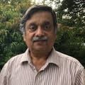
Dr. Suraj Munjal
Ophthalmologist- Retina Surgeon, Cataract Surgeon, Lasik surgeon, Vitreo Retina Surgeon, Eyelid Surgery, Trabeculectomy, Vitreoretinal Surgery, Orbital & Oculoplastic surgery, Refractive surgery, Retina Examination, Glaucoma Evaluation / Treatment, Canaloplasty, Corneal Surgery, Eye Muscle Surgery, Orbital Surgery, Anterior Segment Surgery, Orbital Decompression Surgery for Thyroid Eye Disease, Orbital Trauma, Diabetic Retinopahy, YAG laser Posterior Capsulotomy, Anterior Retinal Cryotherapy, Retinal Surgery, Lens for Keratoconus
The Sight Avenue Eye Hospital Delhi, India
19 Years of Experience
Treating Doctor
Dr. Suresh Ramchandani
Ophthalmologist- Retina Surgeon, UVEA
Center For Sight, Navi Mumbai, Mumbai Mumbai, India
31 Years of Experience
Treating Doctor
Dr. Md. Ali Mosharraf
Ophthalmologist- Cataract Surgeon, Lasik surgeon, Glaucoma Specialist, Ocular Oncology, Cataract treatment, Eye Checkup - General, Phakic OIL Implantation, Cornea Transplant, Treatment for Keratoconus
The Sight Avenue Eye Hospital Delhi, India
19 Years of Experience

Treating Doctor
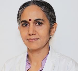
Dr. Rashmi Saraff
Ophthalmologist- Retina Surgeon, Cataract Surgeon, Neuro-ophthalmologist, UVEA
Center For Sight, Rabindra Sadan, Kolkata Kolkata, India
20 Years of Experience
Treating Doctor
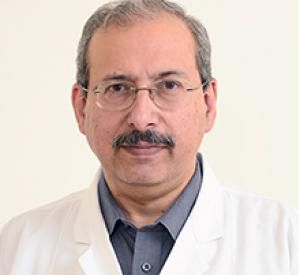
Dr. Dinesh Talwar
Ophthalmologist- Laser Surgeon, Cataract Surgeon, Lasik surgeon, Glaucoma Specialist, Eye Transplant Surgeon, Vitreoretinal Surgery, Strabismus/Squint, Diabetic Retinopahy, YAG laser Posterior Capsulotomy, Cornea Transplant, Anterior Retinal Cryotherapy, Retinal Surgery, Treatment for Keratoconus
Center For Sight New Delhi, India
33 Years of Experience

Treating Doctor
Dr. Vidya Nair Chaudhry
Ophthalmologist- Squint Surgeon, Paediatric Otorhinolaryngology, Oculoplasty, Comprehensive ophthalmology, Comprehensive ophthalmology, Glaucoma and retina
Aakash Healthcare Super Speciality Hospital New Delhi, India
18 Years of Experience
Treating Doctor
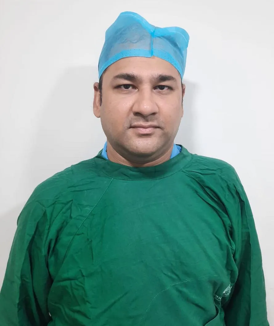
Dr Amanjot Singh
Ophthalmologist- Vitreo Retina Surgeon, Eyelid Surgery, Refractive surgery, Oculoplasty, Glaucoma Evaluation / Treatment, Canaloplasty, Corneal Surgery, Eye Muscle Surgery, Eyelid Surgery, Orbital Decompression Surgery for Thyroid Eye Disease, Orbital Trauma, Refractive surgery
Max Super Speciality Hospital New Delhi, India
26 Years of Experience
Treating Doctor
Dr Tarun Kapur
Ophthalmologist- Eye Surgeon, Cataract Surgeon, Retina Examination, Eye Checkup - General, Glaucoma Evaluation / Treatment
Max Super Speciality Hospital New Delhi, India
29 Years of Experience
Treating Doctor
Dr. Subhrangshu Sengupta
Ophthalmologist- Retina Surgeon, Cataract Surgeon, Lasik surgeon, Neuro-ophthalmologist, Glaucoma Specialist, Refractive surgery, Cataract treatment, UVEA, Refractive surgery
Center For Sight, Rabindra Sadan, Kolkata Kolkata, India
15 Years of Experience

Treating Doctor
Dr. Siddarth Sain
Ophthalmologist- Cataract Surgeon, Phaco Surgeon, Vitreo Retina Surgeon, Cataract treatment, Cataract & Uveitis, Retina Examination, Extracapsular Cataract Extraction, Retinal Surgery
Sharp Sight Eye Hospitals New Delhi, India
13 Years of Experience
Similar Packages
VITREO RETINAL PROCEDURES Pars Plana Viterctomy 25G for Single Eye
India
Start from
$2205 $2095
@ 5%% off