Spinal Tumor Excision With Fixation, Thailand
Spinal Tumor Excision With Fixation
Thailand
-
Our Price USD 9297
-
Hospital Price USD 9786
-
You Save : USD 489
Booking Amount: USD 930. Pay Remaining 90% at the hospital.
Book NowAdditional Credit
Among the important extras we offer as part of the Additional Credit are the following:
-
Site Tourism For The Patient & Attendant
-
Airport Pick & Drop Service
-
Ambulance service at airport
-
Priority appointments with The Doctor
-
Cancel Easily Anytime with Full Refund
-
Room Upgradation
-
Free Online Doctor Consultation Valued at USD 20
-
Free hotel Stay for 5 to 7 days Accordingly
-
Welcome Kit at Arrival
-
Interpreter
-
Medical Visa Assistance
What is Included?
- Doctor consultation charges
- Lab tests and diagnostic charges
- Room charges inside hospital during the procedure
- Surgeon Fee
- Cost of implant
- Nursing charges
- Hospital surgery suite charges
- Anesthesia charges
- Routine medicines and routine consumables (bandages, dressings etc.)
- Food and Beverages inside hospital stay for patient and one attendant.
What is not Included?
- Extra Radiology Investigations
- Healthcare Professionals Charges of other consultations.
- Other Requested Services such as Laundry etc.
- Additional Pharmaceutical Products and Medicines After Discharge from Hospital.
- Management of Conditions Unrelated to Procedures or Pre-Existing.
- The cost of any additional implants will be in addition to the package cost.
Package Description
Spinal Tumor Excision With Fixation
Remove the tumour entirely or a portion of it. Debulking (reduction in size), excise (total removal), and resection are some of the medical words employed (partial removal). These operations assist to alleviate pain and other symptoms by decompressing or relieving pressure on the spinal cord and nerve roots.
Disease Overview:
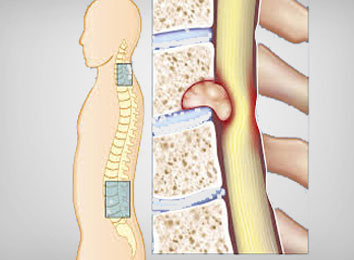
Spinal tumour
A spinal tumour is an abnormal mass of tissue that grows and multiplies uncontrolled within or around the spinal cord and spinal column. The grade, origin, and location of spinal tumours are all categorised. Spinal tumours are classified as either benign (non-cancerous) or malignant (cancerous) (cancerous). Primary spinal tumours arise in the spine or spinal cord, whereas secondary or metastatic spinal tumours arise from cancer that has spread to the spine from another part of the body. The location of the tumour on the spine can also be used to refer to it.
Cervical (neck), thoracic (mid-back), lumbar (lower back), and sacrum are the four fundamental regions (bone at the end of the spine). Additionally, spinal tumours are divided into two groups based on where they are located in the spine: anterior (front) and posterior (back) (back).
Disease Signs and Symptoms:
The most common symptom of both benign and malignant spinal tumours is non-mechanical back discomfort, especially in the middle or lower back. Injury, stress, or physical exercise are not to blame for this back discomfort. The discomfort, on the other hand, may worsen with exercise and is often worst at night while lying down. Even when treated with conservative, nonsurgical approaches that can frequently help ease back pain related to mechanical reasons, discomfort might extend to the hips, legs, feet, or arms and worsen with time. Other indications and symptoms may appear when a tumour develops and compresses the spinal cord, nerve roots, blood vessels, or bones of the spine, depending on the location and kind of tumour
Additional signs and symptoms may include:
- Legs, arms, or chest muscle weakness or loss of feeling
- Neck or back pain
- Valsalva manoeuvre causes pain and/or neurologic symptoms (such as tingling).
- Walking difficulties, which may result in falls
- Sensitivity to pain, heat, and cold is reduced.
- Deficiency in bowel or bladder function
- Depending on which nerves are squeezed, paralysis can develop in differing degrees and in different areas of the body.
- Scoliosis or other deformities of the spine caused by a big and/or destructive tumour
Disease Causes:
The majority of primary spinal tumours have no recognised aetiology. Some of them might be linked to cancer-causing substances. Lymphomas of the spinal cord, which affect lymphocytes (a kind of immune cell), are more frequent in persons who have weakened immune systems. There appears to be a hereditary component to the increased occurrence of spinal tumours in some families.
Primary tumours can emerge from the presence of these two genetic illnesses in a tiny percentage of cases:
Neurofibromatosis 2: Benign tumours may grow in the arachnoid layer of the spinal cord or in the supporting glial cells in this hereditary (genetic) condition. The more prevalent tumours linked with this illness, on the other hand, disrupt the nerves that control hearing and can ultimately lead to hearing loss.
Von Hippel-Lindau disease is a rare multi-system condition that is linked to benign blood vessel tumours (hemangioblastomas) in the brain, retina, and spinal cord, as well as additional malignancies in the kidneys and adrenal glands.
Disease Diagnosis:
The first step in detecting a spinal tumour is a thorough medical evaluation that focuses on back pain and neurological impairments. For a precise and definite diagnosis, radiological examinations are essential.
The structure of the vertebrae and the contour of the joints may be seen using an X-ray, which uses radiation to create a film or image of a region of the body. X-rays of the spine are taken to look for other reasons of pain, such as tumours, infections, fractures, and so on. X-rays, on the other hand, aren't always accurate in detecting cancers.
CT or CAT scan: A diagnostic image formed when a computer scans X-rays, a CT/CAT scan can indicate the shape and size of the spinal canal, as well as its contents and surrounding structures. It's also great for imagining bone structures.
Magnetic resonance imaging (MRI) is a diagnostic examination that uses strong magnets and computer technologies to create three-dimensional pictures of bodily components. An MRI can reveal enlargement, degeneration, and malignancies in the spinal cord, nerve roots, and adjacent regions.
Technectium-99 Bone Scan: This is a diagnostic test that uses Technectium-99. Bone malignancies (such as primary bone tumours of the spine), infection, and disorders involving aberrant bone metabolism can all be detected using this tool.
The above-mentioned radiology examinations yield imaging data that indicate the most likely tumour kind. A biopsy may be required in some circumstances if the diagnosis is ambiguous or if there is a worry about malignancy vs. benign tumour type. A biopsy can also assist establish the kind of cancer, which dictates treatment options if the tumour is malignant.
Staging determines the amount of neoplasms (abnormal tissue) by measuring bone, soft tissue, and spinal canal involvement. For staging purposes, a clinician may request a nuclear-based whole-body scan as well as a CT scan of the lungs and abdomen. A doctor analyses laboratory test results and data from the aforementioned scans to the patient's symptoms to confirm the diagnosis.
Disease Treatment:
Spinal Tumors and Their Treatment
Surgery, radiation, chemotherapy, medicine, or a combination of therapies may be used to treat spinal tumours. The type of spinal tumour and the patient's general health history determine the treatment approach. Our Spinal Tumor Program meets once a week to design and discuss treatment regimens for individual spinal tumour patients.
Treatment for Metastatic Spinal Tumors
Multiple treatment options for metastatic spinal tumours are considered, including surgery, radiation therapy, interventional radiology treatments, and systemic medications including chemotherapy. Many aspects go into determining the appropriate treatment for metastatic spine tumours, and each patient's clinical status must be taken into account. Many factors influence a patient's outcome, including the amount of malignancy, previous cancer therapies, functional status, and the degree of spinal involvement.
Treatment for Primary Spinal Tumors
Primary spinal tumours are treated in a variety of ways, depending on the kind of tumour. An image-guided biopsy is suggested to determine the diagnosis if a primary spinal tumour is suspected. Complete or "en bloc" removal of malignant or locally aggressive tumours such as chordoma or other spinal sarcomas can provide long-term tumour management. En bloc resection is a difficult surgical treatment that entails removing the entire tumour in one piece to prevent the tumour from spreading throughout the process. Other primary spinal tumours can be surgically removed in sections without fear of the tumour spreading.
Treatment for Spinal Cord Tumors
The treatment for spinal cord tumours differs based on the type of tumour and where it is located in the spinal cord. To determine the diagnosis and guide the treatment approach, an image-guided biopsy is done initially when possible. Spinal cord cancers are treated in a variety of ways, including maximising tumour removal with specialist equipment (such as a surgical microscope and intraoperative neuro-monitoring) and/or radiation treatment (e.g., stereotactic radiation procedures or other forms of conformal radiation therapy). Radiation treatment and/or chemotherapy may be considered if the spinal tumour cannot be entirely eliminated.
Information related to Treatment
Package Details
Days in Hospital
3 Days
Days in Hotel
*
10 Days
Room Type
Private
Similar Packages