Craniotomy Surgery cost in India
The cost of Craniotomy Surgery in India ranges
from USD 5700 to USD 11000.
Procedure Description:
Craniotomy Surgery
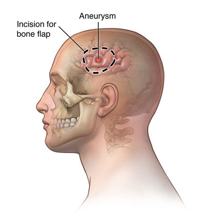
A craniotomy is a type of brain surgery in which a neurosurgeon opens and removes a section of the skull to provide access to the brain. During the same procedure, your surgeon will reattach the piece of your skull. A craniotomy is a serious operation. A surgeon typically considers this sort of treatment after identifying a brain tumor, a life-threatening disease, or a traumatic brain injury.
There are various craniotomy procedures. Their names refer to the place from which your surgeon will remove a piece of your skull to gain access to the brain. Examples of craniotomy operations include:
1- Frontal: The area of your head nearest your hairline.
Temporal: The area of your skull close to your eyes and in front of your ears.
2- Parietal: The top-middle and upper back of the skull.
Pterional (frontotemporal): The side of your skull located behind your temple.
3- Orbitozygomatic: The area of your skull near your eye socket and cheek.
4- Retrosigmoid (keyhole): A tiny incision in the skull below the ear.
Suboccipital: The base of your skull, just above your neck.
Disease Overview:
Brain Tumor
A brain tumor is an abnormal cell growth in the brain's tissues. Brain tumors can be benign (no cancer cells) or malignant (fast-growing cancer cells). Some of them are primary brain tumours, meaning they begin in the brain. Others are metastatic, which means they begin elsewhere in the body and spread to the brain.
As new cells replace old or damaged ones, normal cells proliferate in a regulated manner. Tumor cells multiply uncontrolled for reasons that are unknown.
A primary brain tumor is a benign tumor that begins in the brain and seldom spreads to other regions of the body. Primary brain tumors can be either benign or cancerous.
A benign brain tumor develops slowly, has well-defined borders, and spreads only infrequently. Benign tumors can be life threatening if they are placed in a key region, despite the fact that their cells are not cancerous.
A malignant brain tumor spreads to neighboring brain regions, develops swiftly, and has irregular borders. Malignant brain tumors, despite their common name, do not meet the criteria of cancer since they do not spread to organs outside of the brain and spine.
Metastatic (secondary) brain tumors start out as cancer in another part of the body and then spread to the brain. When cancer cells are transported through the bloodstream, they develop tumors. Lung and breast cancers are the most prevalent malignancies that spread to the brain.
A brain tumor, whether benign, malignant, or metastatic, can all be life-threatening. The brain can't expand to make place for a growing mass since it's encased in a bony skull. The tumour compresses and displaces normal brain tissue as a result.
Some brain tumors cause the cerebrospinal fluid (CSF) that circulates around and through the brain to become clogged. This obstruction raises intracranial pressure and can cause the ventricles to expand (hydrocephalus). Swelling is a symptom of certain brain tumors (edema). The "mass effect" is caused by the size, pressure, and swelling of the body, which causes many of the symptoms.
Disease Signs and Symptoms:
Tumors can cause damage to the brain by killing healthy tissue, squeezing healthy tissue, or raising intracranial pressure. The kind, size, and location of the tumour in the brain all influence the symptoms. Symptoms in general include:
- Seizures with headaches that seem to get worse in the morning
- stumbling, dizziness, and walking difficulties
- issues with speech (e.g., difficulty finding the right word)
- irregular eye movements, visual difficulties
- Increased intracranial pressure due to weakness on one side of the body produces sleepiness, headaches, nausea and vomiting, and slow reactions.
The following are examples of specific symptoms:
Behavioral and emotional problems; poor judgement, motivation, or inhibition; decreased sense of smell or visual loss; paralysis on one side of the body; lower mental ability and memory loss are all possible adverse effects of frontal lobe tumours.
Parietal lobe tumours can cause difficulty with speaking, writing, drawing, and naming, as well as lack of recognition, spatial impairments, and eye-hand coordination.
Vision loss in one or both eyes, visual field cuts, fuzzy vision, illusions, and hallucinations are all possible symptoms of occipital lobe tumours.
Temporal lobe tumours can cause issues with speaking and interpreting language, as well as short- and long-term memory.
aggressiveness on the rise
Behavioral and emotional problems, trouble speaking and eating, tiredness, hearing loss, muscular weakness on one side of the face (e.g., head tilt, crooked grin), uncoordinated walking, drooping eyelid or double vision, and vomiting are all symptoms of brainstem tumours.
Increased hormone secretion (Cushing's Disease, acromegaly), cessation of menstruation, irregular milk secretion, and diminished libido are all possible side effects of pituitary gland tumours.
Disease Causes:
hereditary illnesses, such as neurofibromatosis, extended exposure to pesticides, industrial solvents, and other toxins cancer elsewhere in the body
Country wise cost comparison for Craniotomy Surgery:
| Country | Cost |
|---|---|
| India | $6120 |
| Thailand | $21267 |
| Iran | $4980 |
| Singapore | $49219 |
Treatment and Cost
15
Total Days
In Country
- 5 Day in Hospital
- 2 No. Travelers
- 10 Days Outside Hospital
Treatment cost starts from
$6800
Popular Hospital & Clinic
Featured Hospital
0 Hospitals
Related Packages
More Related Information
Some of the top rated hospitals are:
- Turkey
- Kolan International Hospital, Sisli
- Istinye University Bahcesehir Liv Hospital
- Istinye University Medical Park Gaziosmanpasa Hospital
- I.A.U VM Medical Park Florya Hospital
- Altinbas University Medical Park Bahcelievler Hospital
- Medical Park Antalya Hospital
- Medical Park Tarsus Hospital, Mersin
- Thailand
- Bangpakok 9 International Hospital
- Bumrungrad International Hospital
- Bangkok Hospital
- Bangkok International Hospital
- Samitivej Hospital
- BNH Hospital
- Aek Udon International Hospital
- Phuket International Hospital
- Bangkok Christian Hospital
- Thonburi Hospital
- Kasemrad Hospital Sriburin
- Saudi Arabia