Gama Knife for Brain Tumor cost in India
The cost of Gama Knife for Brain Tumor in
India ranges from USD 6800 to USD 11000
Gama Knife for Brain Tumor:
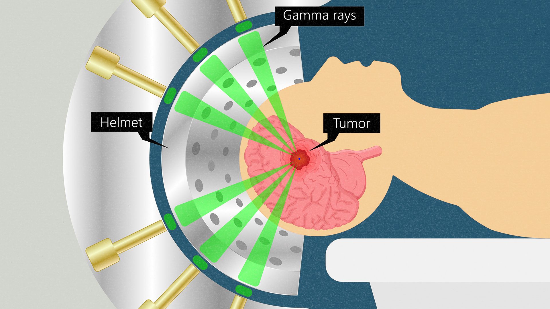
Gamma Knife stereotactic radiosurgery technology delivers a precise dosage of radiation to a target using numerous tiny gamma rays. Gamma Knife radiosurgery is a type of radiation therapy for the treatment of brain tumours, vascular malformations, and other abnormalities.
Disease Overview:
Brain tumors
A brain tumour is an abnormal cell growth in the brain's tissues. Brain tumours can be benign (no cancer cells) or malignant (fast-growing cancer cells). Some of them are primary brain tumours, meaning they begin in the brain. Others are metastatic, which means they begin elsewhere in the body and spread to the brain.
As new cells replace old or damaged ones, normal cells proliferate in a regulated manner. Tumor cells multiply uncontrolled for reasons that are unknown.
A primary brain tumour is a benign tumour that begins in the brain and seldom spreads to other regions of the body. Primary brain tumours can be either benign or cancerous.
A benign brain tumour develops slowly, has well-defined borders, and spreads only infrequently. Benign tumours can be life threatening if they are placed in a key region, despite the fact that their cells are not cancerous.
A malignant brain tumour spreads to neighbouring brain regions, develops swiftly, and has irregular borders. Malignant brain tumours, despite their common name, do not meet the criteria of cancer since they do not spread to organs outside of the brain and spine.
Metastatic (secondary) brain tumours start out as cancer in another part of the body and then spread to the brain. When cancer cells are transported through the bloodstream, they develop tumours. Lung and breast cancers are the most prevalent malignancies that spread to the brain.
A brain tumour, whether benign, malignant, or metastatic, can all be life-threatening. The brain can't expand to make place for a growing mass since it's encased in a bony skull. The tumour compresses and displaces normal brain tissue as a result.
Some brain tumours cause the cerebrospinal fluid (CSF) that circulates around and through the brain to become clogged. This obstruction raises intracranial pressure and can cause the ventricles to expand (hydrocephalus). Swelling is a symptom of certain brain tumours (edema). The "mass effect" is caused by the size, pressure, and swelling of the body, which causes many of the symptoms.
Disease Signs and Symptoms:
Medical research has yet to discover what causes brain tumours or how to avoid primary brain tumours. Those with the following conditions are at the highest risk of developing a brain tumour:
hereditary illnesses, such as neurofibromatosis, extended exposure to pesticides, industrial solvents, and other toxins cancer elsewhere in the body
Disease Causes:
Tumors can cause damage to the brain by killing healthy tissue, squeezing healthy tissue, or raising intracranial pressure. The kind, size, and location of the tumour in the brain all influence the symptoms. Symptoms in general include:
Seizures with headaches that seem to get worse in the morning
stumbling, dizziness, and walking difficulties
issues with speech (e.g., difficulty finding the right word)
irregular eye movements, visual difficulties
Increased intracranial pressure due to weakness on one side of the body produces sleepiness, headaches, nausea and vomiting, and slow reactions.
The following are examples of specific symptoms:
Behavioral and emotional problems; poor judgement, motivation, or inhibition; decreased sense of smell or visual loss; paralysis on one side of the body; lower mental ability and memory loss are all possible adverse effects of frontal lobe tumours.
Parietal lobe tumours can cause difficulty with speaking, writing, drawing, and naming, as well as lack of recognition, spatial impairments, and eye-hand coordination.
Vision loss in one or both eyes, visual field cuts, fuzzy vision, illusions, and hallucinations are all possible symptoms of occipital lobe tumours.
Temporal lobe tumours can cause issues with speaking and interpreting language, as well as short- and long-term memory.
aggressiveness on the rise
Behavioral and emotional problems, trouble speaking and eating, tiredness, hearing loss, muscular weakness on one side of the face (e.g., head tilt, crooked grin), uncoordinated walking, drooping eyelid or double vision, and vomiting are all symptoms of brainstem tumours.
Increased hormone secretion (Cushing's Disease, acromegaly), cessation of menstruation, irregular milk secretion, and diminished libido are all possible side effects of pituitary gland tumours.
Disease Diagnosis:
First, the doctor will gather information about your personal and family medical history before doing a thorough physical examination. A neurological exam is performed in addition to a routine physical examination to assess mental status and memory, cranial nerve function (sight, hearing, smell, tongue and facial movement), muscular strength, coordination, reflexes, and pain response. Additional testing may include the following:
- Hearing loss caused by tumours near the cochlear nerve can be detected by audiometry, a hearing test conducted by an audiologist (e.g., acoustic neuroma).
- An endocrine examination detects abnormal hormone levels produced by pituitary tumours (e.g., Cushing's Disease) by measuring hormone levels in your blood or urine.
- A neuro-ophthalmologist performs a visual field acuity test to discover vision loss and missing spots in your range of vision.
- A lumbar puncture (spinal tap) can be used to look for tumour cells, proteins, infection, and blood in the cerebrospinal fluid.
- Tests of imaging
An X-ray beam and a computer are used to study anatomical features during a computed tomography (CT) scan. It takes pictures of each slice of the brain as it cuts it layer by layer. You may be given a dye (contrast agent) to inject into your bloodstream. CT scans are particularly useful for examining changes in bony structures.
A magnetic field and radiofrequency waves are used in an MRI scan to provide a thorough image of the brain's soft tissues. It sees the brain in three dimensions, in cross-sections that may be taken from the side or from the top. You may be given a dye (contrast agent) to inject into your bloodstream. The use of MRI to assess brain lesions and their consequences on the surrounding brain is extremely beneficial.
Disease Treatment:
The kind, grade, size, and location of the tumour, as well as whether it has spread and your age and overall health, all influence treatment options. The purpose of therapy may be curative or symptomatic relief (palliative care). Treatments are frequently used in tandem with one another. The purpose of surgery is to remove all or as much of the tumour as feasible in order to reduce the risk of recurrence. Tumors that cannot be removed by surgery are treated with radiation treatment and chemotherapy. For instance, surgery may remove the majority of the tumour, leaving only a little bit of tumour near a crucial structure to be treated with radiation later.
Observation
Observation is sometimes the best medicine. For example, benign, slow-growing tumours that are tiny and have few symptoms can be monitored with annual MRI scans until they become large enough to require surgery. For persons who are elderly or have other health problems, observation may be the best option.
Surgery
For brain tumours that can be accessed without inflicting substantial harm to essential regions of the brain, surgery is the therapy of choice. Surgery can aid in the refinement of the diagnosis, the removal of as much of the tumour as feasible, and the relief of pressure within the skull. A craniotomy is performed by a neurosurgeon to open the skull and remove the tumour. If the tumour is near vital parts of the brain, just a portion of it may be removed. Even if only a portion of the tumour is removed, symptoms can be relieved. On the residual tumour cells, radiation or chemotherapy may be utilised.
The surgeon's ability to precisely locate the tumour, define the tumor's borders, avoid injury to vital brain areas, and confirm the amount of tumour removal while in the operating room has improved thanks to image-guided surgery technologies, tumour fluorescence, intraoperative MRI/CT, and functional brain mapping.
Laser Interstitial Thermal Therapy is a type of interstitial thermal therapy that uses laser
Laser ablation is a minimally invasive therapy in which heat is sent from the inside out to "cook" brain tumours. Through a burr hole in the skull, a probe is introduced into the tumour. Real-time MRI is used to guide the laser catheter.
Radiation
Radiation therapy is a treatment for brain cancers that involves high-energy radiation that are carefully regulated. Radiation destroys the DNA in cells, preventing them from dividing and growing. The advantages of radiation are not instantaneous, but they develop over time. Radiation is more effective against aggressive cancers because their cells proliferate quickly. The aberrant cells die and the tumour shrinks over time. Benign tumours take months to manifest because their cells proliferate slowly.
In a single session, stereotactic radiosurgery (SRS) provides a high dosage of radiation. The subject is immobilised using frames and masks.
Fractionated radiotherapy gives smaller radiation doses over a longer period of time. Patients must return every day for many weeks to obtain the full dose of radiation.
Proton beam treatment targets the tumour at a specified depth using accelerated proton energy. The beam of radiation does not extend beyond the tumour.
The radiation dosage is delivered to the entire brain using whole brain radiotherapy (WBRT). It has the potential to treat a variety of brain cancers and metastases.
Chemotherapy
Chemotherapy medications function by interfering with the division of cells. Chemotherapy causes aberrant cells to die and the tumour to shrink over time. Normal cells can also be damaged by this therapy, although they are more capable of self-repair than aberrant cells. Treatment is given in cycles, with periods of rest in between to allow the body to regenerate healthy cells.
Country wise cost comparison for Gama Knife for Brain Tumor:
| Country | Cost |
|---|---|
| India | $6250 |
| Turkey | $9787 |
| Singapore | $58046 |
Treatment and Cost
15
Total Days
In Country
- 1 Day in Hospital
- 2 No. Travelers
- 14 Days Outside Hospital
Treatment cost starts from
$6944
Popular Hospital & Clinic
Featured Hospital
10 Hospitals
Types of Gama Knife for Brain Tumor in Fortis Memorial Research Institute and its associated cost
| Treatment Option | Approximate Cost Range (USD) |
|---|---|
| No Treatment option added | |
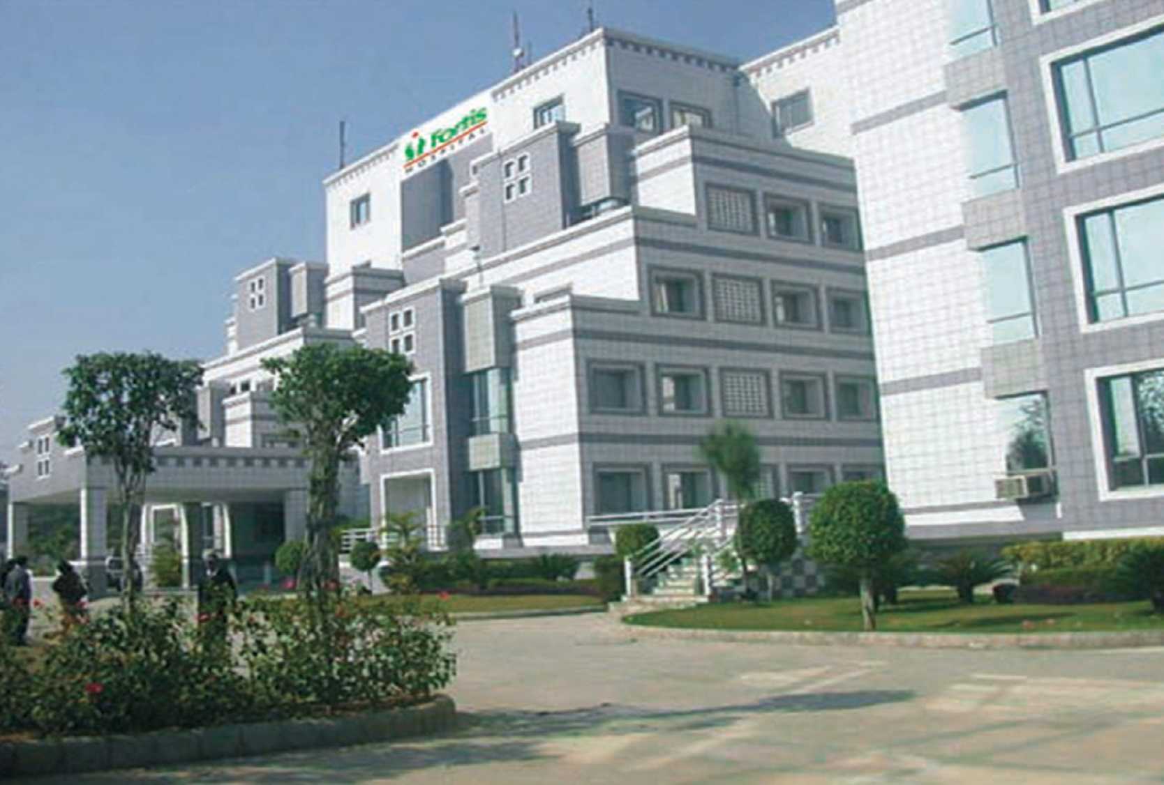
- Address: Sector - 44, Opp. HUDA City Center,Gurgaon, Haryana - 122002, India
- Facilities related to Fortis Memorial Research Institute: Private Rooms, Translator, Nursery / Nanny Services, Airport Pick up, Personal Assistance / Concierge, Free Wifi, Local Tourism Options, International Cuisine, Phone in Room, Private Driver / Limousine Services, Post operative followup, Mobility Accessible Rooms, Online Doctor Consultation, Air Ambulance, Religious Facilities, Rehabilitation, Cafe, TV in room, Car Hire, Health Insurance Coordination,
50
DOCTORS IN 35 SPECIALITIES
20+
FACILITIES & AMENITIES
Types of Gama Knife for Brain Tumor in CK Birla Hospital, Gurgaon and its associated cost
| Treatment Option | Approximate Cost Range (USD) |
|---|---|
| No Treatment option added | |
- Address: Block J, CK Birla Hospital, Nirvana Central Rd, Mayfield Garden, Sector 51, Gurugram, Haryana 122018
- Facilities related to CK Birla Hospital, Gurgaon: The hospital has following facilities: Patient Rooms: Presidential suite, suites, junior suites, deluxe single and shared rooms 4 state-of-the-art modular OTs Critical Care Facilities Labour delivery rooms (LDR) with piped Entonox for pain management North India’s only water-birthing facility Advanced IVF laboratory Chemo Daycare center Physiotherapy Centre 24x7 Radiology - including 4D ultrasound, X-ray, digital mammography, BMD, ECHO 24x7 Pathology including advanced genetic testing 24x7 Emergency 24x7 Pharmacy Studio for fitness, antenatal and post-natal classes & to host medical and educational workshops Café serving healthy & nutritious food and drinks
13
DOCTORS IN 21 SPECIALITIES
20+
FACILITIES & AMENITIES
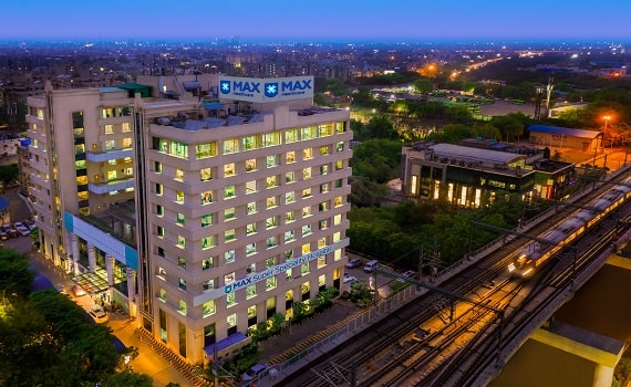
Types of Gama Knife for Brain Tumor in BLK-Max Super Speciality Hospital and its associated cost
| Treatment Option | Approximate Cost Range (USD) |
|---|---|
| No Treatment option added | |
- Address: Pusa Road, New Delhi-110005
- Facilities related to BLK-Max Super Speciality Hospital: Private Rooms, Translator, Nursery / Nanny Services, Personal Assistance / Concierge, Free Wifi, International Cuisine, Phone in Room, Private Driver / Limousine Services, Post operative follow-up, Mobility Accessible Rooms, Rehabilitation, Cafe, TV in room, Car Hire, Health Insurance Coordination
17
DOCTORS IN 33 SPECIALITIES
20+
FACILITIES & AMENITIES
Types of Gama Knife for Brain Tumor in Max Super Speciality Hospital and its associated cost
| Treatment Option | Approximate Cost Range (USD) |
|---|---|
| No Treatment option added | |
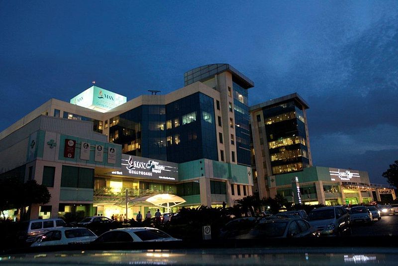
- Address: Max Super Speciality Hospital No. 1, 2, Press Enclave Road, Mandir Marg, Saket Institutional Area, Saket, New Delhi, Delhi, 110017, India
- Facilities related to Max Super Speciality Hospital:
53
DOCTORS IN 34 SPECIALITIES
20+
FACILITIES & AMENITIES
Types of Gama Knife for Brain Tumor in Fortis Escorts Heart Institute and its associated cost
| Treatment Option | Approximate Cost Range (USD) |
|---|---|
| No Treatment option added | |
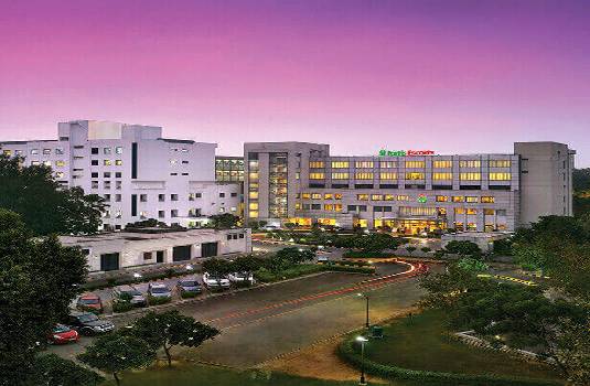
- Address: Okhla Road,New Delhi - 110 025 (INDIA)
- Facilities related to Fortis Escorts Heart Institute: Private Rooms, Translator, Nursery / Nanny Services, Personal Assistance / Concierge, Free Wifi, International Cuisine, Phone in Room, Private Driver / Limousine Services, Post operative follow-up, Mobility Accessible Rooms, Rehabilitation, Cafe, TV in room, Car Hire, Health Insurance Coordination
19
DOCTORS IN 33 SPECIALITIES
20+
FACILITIES & AMENITIES
Types of Gama Knife for Brain Tumor in PSRI Hospital and its associated cost
| Treatment Option | Approximate Cost Range (USD) |
|---|---|
| No Treatment option added | |
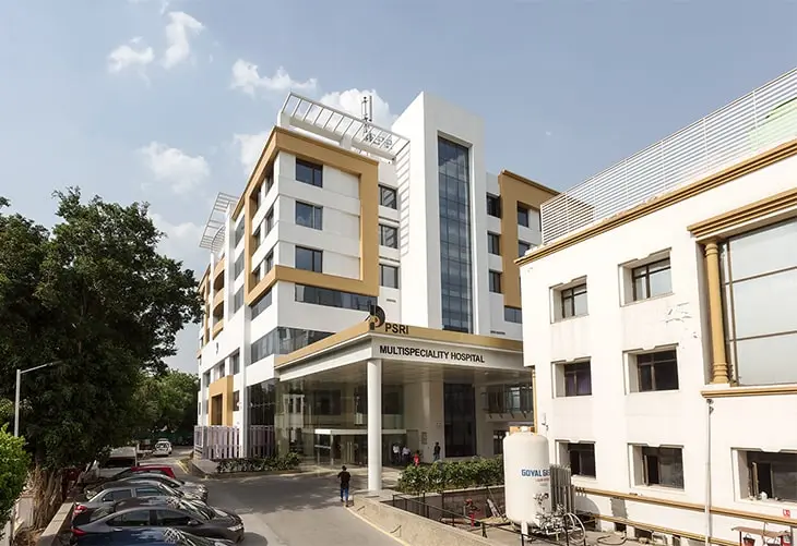
- Address: Press Enclave Marg, J Pocket, Phase II, Sheikh Sarai, New Delhi, Delhi 110017
- Facilities related to PSRI Hospital: Private Rooms, Translator, Nursery / Nanny Services, Personal Assistance / Concierge, Free Wifi, International Cuisine, Phone in Room, Private Driver / Limousine Services, Post operative follow-up, Mobility Accessible Rooms, Rehabilitation, Cafe, TV in room, Car Hire, Health Insurance Coordination
8
DOCTORS IN 33 SPECIALITIES
20+
FACILITIES & AMENITIES
Types of Gama Knife for Brain Tumor in Fortis Flt. Lt. Rajan Dhall Hospital, Vasant Kunj, Delhi and its associated cost
| Treatment Option | Approximate Cost Range (USD) |
|---|---|
| No Treatment option added | |
- Address: Fortis Flt. Lt. Rajan Dhall Hospital, Aruna Asaf Ali Marg, Pocket 1, Sector B, Vasant Kunj, New Delhi, Delhi 110070
- Facilities related to Fortis Flt. Lt. Rajan Dhall Hospital, Vasant Kunj, Delhi: Private Rooms, Translator, Nursery / Nanny Services, Personal Assistance / Concierge, Free Wifi, International Cuisine, Phone in Room, Private Driver / Limousine Services, Post operative follow-up, Mobility Accessible Rooms, Rehabilitation, Cafe, TV in room, Car Hire, Health Insurance Coordination
46
DOCTORS IN 34 SPECIALITIES
20+
FACILITIES & AMENITIES
Types of Gama Knife for Brain Tumor in MAX Super Speciality hospital, Patpadganj Delhi and its associated cost
| Treatment Option | Approximate Cost Range (USD) |
|---|---|
| No Treatment option added | |
- Address: 108A, Indraprasth Extension, Patpadganj, New Delhi- 110092, India
- Facilities related to MAX Super Speciality hospital, Patpadganj Delhi: Private Rooms, Translator, Nursery / Nanny Services, Personal Assistance / Concierge, Free Wifi, International Cuisine, Phone in Room, Private Driver / Limousine Services, Post operative follow-up, Mobility Accessible Rooms, Rehabilitation, Cafe, TV in room, Car Hire, Health Insurance Coordination
52
DOCTORS IN 33 SPECIALITIES
20+
FACILITIES & AMENITIES
Types of Gama Knife for Brain Tumor in Medanta-The Medicity, Gurgaon and its associated cost
| Treatment Option | Approximate Cost Range (USD) |
|---|---|
| No Treatment option added | |
- Address: CH Baktawar Singh Road, Sector 38, Gurugram, Haryana 122001
- Facilities related to Medanta-The Medicity, Gurgaon: TV in room Private rooms, Free Wifi, Phone in Room, Mobility accessible rooms, Family accommodation, Laundry, Welcome Safe in the room, Nursery / Nanny services. Dry cleaning, Personal assistance / Concierge Religious facilities, Fitness Spa and wellness Café, Business Centre, Shop, Dedicated smoking areas, Beauty Salon, Special offer for group stays, Parking available, Health insurance coordination, Medical travel insurance, Foreign currency exchange, ATM, Credit Card, Debit Card, Net banking, Diet on Request, Restaurant, International Cuisine, Treatment Related Medical records transfer, Online doctor consultation, Rehabilitation, Pharmacy, Document legalization, Post operative follow-up, Language Interpreter, Translation services, Transportation, Airport pickup, Local tourism options, Local transportation booking, Visa / Travel office, Car Hire, Private driver / Limousine services, Air ambulance
52
DOCTORS IN 33 SPECIALITIES
20+
FACILITIES & AMENITIES
Types of Gama Knife for Brain Tumor in Indraprastha Apollo Hospitals, New Delhi and its associated cost
| Treatment Option | Approximate Cost Range (USD) |
|---|---|
| No Treatment option added | |
- Address: Mathura Rd, Jasola Vihar, New Delhi, Delhi 110076
- Facilities related to Indraprastha Apollo Hospitals, New Delhi: Private Rooms, Translator, Nursery / Nanny Services, Personal Assistance / Concierge, Free Wifi, International Cuisine, Phone in Room, Private Driver / Limousine Services, Post operative follow-up, Mobility Accessible Rooms, Rehabilitation, Cafe, TV in room, Car Hire, Health Insurance Coordination
37
DOCTORS IN 33 SPECIALITIES
20+
FACILITIES & AMENITIES
Related Packages
More Related Information
Some of the top rated doctors are:
- Saudi Arabia
Some of the top rated hospitals are:
Frequently Asked Questions
The cost of gamma knife radiosurgery in India varies depending on the hospital. The best gamma-knife radiosurgery hospitals in India pay for all of the candidate's pre-surgery investigation costs. The total cost of the Gamma Knife Radiosurgery package includes the cost of the procedure, medications, consumables, and investigations. New discoveries, post-operative problems, and a delayed recovery could affect the overall cost of gamma knife radiosurgery in India.
The starting cost of Gamma Knife Radiosurgery in India is approximately USD $5,500. Gamma Knife Radiosurgery is available at numerous facilities in India that are JCI and NABH certified.
In India, there are numerous hospitals that use Gamma Knife Radiosurgery. The following hospitals are among the most well-known in India for Gamma Knife Radiosurgery:
1-Artemis Health Institute
2-Fortis Hospital
3-Medanta - The Medicity
4-Apollo Hospital
5-Max Super Speciality Hospital, Patparganj
6-Sterling Wockhardt Hospital
7-The Madras Institute of Orthopedics and Traumatology (MIOT)
8-Max Super Specialty Hospital, Vaishali
9-Manipal Hospitals, Goa
10-Dr. Rela Institute and Medical Centre
The following are some of the top Indian cities where Gamma Knife Radiosurgery is available:
New Delhi, Chennai, Mumbai, and Ghaziabad.
In addition to the cost of the Gamma Knife Radiosurgery, the patient can also be responsible for other daily costs like meals and lodging after discharge. In India, the daily additional costs per person are approximately $25 USD.
Following Gamma Knife Radiosurgery, the patient is expected to remain in the hospital for recuperation and observation for approximately one day. It's crucial that the patient recovers appropriately and feels comfortable following the procedure within this time range. It is established through a battery of tests that the patient is doing well following surgery and can be released.

