Vitrectomy cost in India
The cost of Vitrectomy in India ranges
from USD 1000 to USD 4000
Procedure Overview:
Vitrectomy
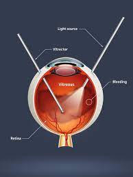
A vitrectomy is a procedure where the vitreous humor in the eye is removed whole or in part. Small amounts of the vitreous humor from the front structures of the eye are removed during an anterior vitrectomy; this is typically done because the vitreous humor has become entangled with an intraocular lens or other structures.
Disease Overview:
Retinal detachment is a condition in which a thin layer of tissue at the back of the eye (the retina) slips away from its usual position. Retinal detachment occurs when the layer of blood vessels that supplies oxygen and sustenance to the retina separates from the retinal cells.
The longer you wait to treat a retinal detachment, the more likely you are to lose vision in the afflicted eye permanently.
The abrupt emergence of floaters and flashes, as well as impaired vision, are all possible symptoms of retinal detachment. Contacting an ophthalmologist (eye doctor) as soon as possible will help you save your vision.
Disease Signs and Symptoms:
The process of retinal detachment is painless. However, there are virtually always warning signals before it develops or has progressed, such as:
- The unexpected emergence of a large number of floaters – little specks that appear to wander across your field of vision
- Light flashes in one or both eyes (photopsia)
- Vision is hazy
- Reduced side (peripheral) vision with time
- Over your visual field, there is a curtain-like shadow.
Disease Causes:
Retinal detachment may be classified into three types:
Rhegmatogenous: These are the most prevalent forms of retinal detachments. A hole or tear in the retina allows fluid to travel through and gather behind the retina, forcing the retina away from the underlying tissues, causing rhegmatogenous detachments. The portions of the retina that detach lose blood flow and stop operating, resulting in visual loss.
Tractional. When scar tissue forms on the surface of the retina, the retina pulls away from the back of the eye, producing detachment. People with poorly managed diabetes or other diseases are more likely to develop tractional separation.
Exudative. Fluid collects behind the retina in this form of detachment, but there are no holes or tears in the retina. Age-related macular degeneration, eye injuries, malignancies, and inflammatory illnesses can all produce exudative detachment.
Risk Factors
The following variables raise your chances of developing a retinal detachment:
- Retinal detachment is more likely in adults over 50 years old.
- One eye had a previous retinal detachment.
- Retinal detachment in the family
- Nearsightedness to the extreme (myopia)
- Previous eye surgery, such as cataract removal, is a good example.
- A previous eye damage was serious.
- Retinoschisis, uveitis, or weakening of the peripheral retina are examples of previous eye diseases or disorders (lattice degeneration)
Disease Diagnosis:
To diagnose retinal detachment, your doctor may utilise the following tests, tools, and procedures:
Examination of the retina. The doctor may check the back of your eye, including the retina, with a tool that includes a powerful light and specific lenses. This sort of gadget allows your doctor to see any retinal holes, tears, or detachments by providing a very detailed image of your whole eye.
Ultrasound imaging is a type of imaging that uses sound waves to create If there has been bleeding in the eye and it is difficult to view your retina, your doctor may utilise this test.
Even if you only have symptoms in one eye, your doctor would most likely examine both.
If your doctor does not find a tear at this appointment, he or she may ask you to return after a few weeks to ensure that your eye has not developed a delayed tear as a consequence of the same vitreous separation. It's also critical to contact your doctor as soon as you notice any new symptoms.
Disease Treatment:
A retinal tear, perforation, or detachment is usually invariably repaired with surgery. There are a variety of strategies to choose from. Inquire about the risks and advantages of your treatment choices with your ophthalmologist. You may decide which surgery or combination of procedures is ideal for you by working together.
Tears in the retina
Your eye surgeon may recommend one of the following procedures to prevent retinal detachment and maintain vision if a retinal tear or hole hasn't proceeded to separation.
Laser surgery is a procedure that involves the use of (photocoagulation). A laser beam is sent into the eye through the pupil by the surgeon. The laser creates scarring around the retinal tear, which "welds" the retina to the underlying tissue.
The process of freezing (cryopexy). After numbing your eye with a local anaesthetic, the surgeon uses a freezing probe to freeze the outer surface of your eye exactly above the tear. The freezing creates a scar that aids in the retina's attachment to the eye wall.
Both of these operations are performed in a hospital setting. Following your treatment, you'll probably be recommended to avoid activities that might jolt your eyes, such as running, for a few weeks.
If your retina has detached, you'll require surgery to restore it as soon as possible after receiving your diagnosis. The type of surgery recommended by your surgeon will be determined by a number of criteria, including the severity of the separation.
Air or gas is injected into your eye. The surgeon injects a bubble of air or gas into the centre of the eye in a technique known as pneumatic retinopexy (RET-ih-no-pek-see) (the vitreous cavity). The bubble, when appropriately positioned, presses the portion of the retina containing the hole or holes against the eye's wall, preventing fluid from flowing into the space behind the retina. During the operation, your doctor may employ cryopexy to heal the retinal break.
Fluid that has accumulated underneath the retina is absorbed by itself, allowing the retina to cling to the eye's wall. To keep the bubble in the appropriate position, you may need to hold your head in a specific posture for several days. The bubble will ultimately dissolve on its own.
The surface of your eye is indented. The surgeon sews (sutures) a piece of silicone material to the white of your eye (sclera) over the afflicted region in a process known as scleral buckling. This treatment indents the eye's wall, relieving part of the tension exerted on the retina by the vitreous.
Your surgeon may make a scleral buckle that encircles your entire eye like a belt if you have multiple tears or holes or an extensive separation. The buckle is positioned in such a manner that it does not obstruct your vision, and it typically stays in place indefinitely.
The fluid in the eye is drained and replaced. The vitreous, as well as any tissue tugging on the retina, is removed during this treatment, known as vitrectomy. The vitreous area is then injected with air, gas, or silicone oil to assist flatten the retina.
The air, gas, or liquid will eventually be absorbed, and the vitreous area will be replenished with bodily fluid. Silicone oil may be surgically removed months later if it was used.
A scleral buckling treatment can be done with a vitrectomy.
It may take several months for your eyesight to improve after surgery. For a successful therapy, you may require a second operation. Some people never fully recover their vision.
Country wise cost comparison for Vitrectomy:
| Country | Cost |
|---|---|
| India | $1403 |
| Iran | $5491 |
Treatment and Cost
15
Total Days
In Country
- 1 Day in Hospital
- 2 No. Travelers
- 14 Days Outside Hospital
Treatment cost starts from
$1477
Popular Hospital & Clinic
Featured Hospital
0 Hospitals
Related Packages
More Related Information
Some of the top rated hospitals are:
- Iran
- Hasheminejad Kidney Center (HKC) Tehran, Iran
- Kasra Hospital Tehran, Iran
- Bahman Hospital Tehran, Iran
- Atieh Hospital Tehran, Iran
- Erfan Hospital Tehran, Iran
- Nikan Hospital Tehran, Iran
- United Arab Emirates
- Burjeel Hospital, Abu Dhabi
- New Hope IVF Gynaecology & Fertility Hospital, Sharjah
- Iranian Hospital, Dubai
- Kings College Hospital Dubai
- Zulekha Hospital Sharjah
- Burjeel Hospital for Advanced Surgery Dubai
- Burjeel Medical City, Abu Dhabi
- NMC Royal Hospital, Khalifa City, Abu Dhabi
- NMC Royal Hospital Sharjah
- AL NOOR HOSPITAL, Abu Dhabi
- Al Zahra hospital, Dubai
- NMC Specialty Hospital, Al Nahda, Dubai
- United Kingdom
- Cancer Centre London
- The Highgate Hospital
- The Holly Hospital
- The Parkside Hospital
- Circle Reading Hospital
- Shirley Oaks Hospital
- St Edmunds Hospital
- The London Clinic
- Woodlands Hospital
- The Christie NHS Foundation Trust
- Royal Marsden Hospital
- Queen Elizabeth Hospital Birmingham
- London Bridge Hospital, HCA Healthcare