Brain Tumor Surgery cost in United
Procedure Description:
Brain Tumor Surgery:
The kind, grade, size, and location of the tumour, as well as whether it has spread and your age and overall health, all influence treatment options. The purpose of therapy may be curative or symptomatic relief (palliative care). Treatments are frequently used in tandem with one another. The purpose of surgery is to remove all or as much of the tumour as feasible in order to reduce the risk of recurrence. Tumors that cannot be removed by surgery are treated with radiation treatment and chemotherapy. For instance, surgery may remove the majority of the tumour, leaving only a little bit of tumour near a crucial structure to be treated with radiation later.
Observation
Observation is sometimes the best medicine. For example, benign, slow-growing tumours that are tiny and have few symptoms can be monitored with annual MRI scans until they become large enough to require surgery. For persons who are elderly or have other health problems, observation may be the best option.
Surgery
For brain tumours that can be accessed without inflicting substantial harm to essential regions of the brain, surgery is the therapy of choice. Surgery can aid in the refinement of the diagnosis, the removal of as much of the tumour as feasible, and the relief of pressure within the skull. A craniotomy is performed by a neurosurgeon to open the skull and remove the tumour. If the tumour is near vital parts of the brain, just a portion of it may be removed. Even if only a portion of the tumour is removed, symptoms can be relieved. On the residual tumour cells, radiation or chemotherapy may be utilised.
The surgeon's ability to precisely locate the tumour, define the tumor's borders, avoid injury to vital brain areas, and confirm the amount of tumour removal while in the operating room has improved thanks to image-guided surgery technologies, tumour fluorescence, intraoperative MRI/CT, and functional brain mapping.
Laser Interstitial Thermal Therapy is a type of interstitial thermal therapy that uses laser
Laser ablation is a minimally invasive therapy in which heat is sent from the inside out to "cook" brain tumours. Through a burr hole in the skull, a probe is introduced into the tumour. Real-time MRI is used to guide the laser catheter.
Radiation
Radiation therapy is a treatment for brain cancers that involves high-energy radiation that are carefully regulated. Radiation destroys the DNA in cells, preventing them from dividing and growing. The advantages of radiation are not instantaneous, but they develop over time. Radiation is more effective against aggressive cancers because their cells proliferate quickly. The aberrant cells die and the tumour shrinks over time. Benign tumours take months to manifest because their cells proliferate slowly.
In a single session, stereotactic radiosurgery (SRS) provides a high dosage of radiation. The subject is immobilised using frames and masks.
Fractionated radiotherapy gives smaller radiation doses over a longer period of time. Patients must return every day for many weeks to obtain the full dose of radiation.
Proton beam treatment targets the tumour at a specified depth using accelerated proton energy. The beam of radiation does not extend beyond the tumour.
The radiation dosage is delivered to the entire brain using whole brain radiotherapy (WBRT). It has the potential to treat a variety of brain cancers and metastases.
Chemotherapy: Chemotherapy medications function by interfering with the division of cells. Chemotherapy causes aberrant cells to die and the tumour to shrink over time. Normal cells can also be damaged by this therapy, although they are more capable of self-repair than aberrant cells. Treatment is given in cycles, with periods of rest in between to allow the body to regenerate healthy cells.
ZAP-X Gyroscopic Radiosurgery: ZapX brain surgery is a state-of-the-art technique designed to accurately identify and manage brain tumors. ZAP-X employs a novel self-shielded, gyroscopic linear accelerator architecture in contrast to more traditional techniques. As a result, it may target tumors from every direction surrounding the head, increasing its effectiveness and precision.
ZAP-X has a high success rate and little long-term negative effects, making it a viable treatment for a variety of illnesses. Because of ZAP-X's accuracy, specific treatments for a range of brain-related disorders are available. ZAP-X aids in:
1. Benign Brain Tumors: Assists in the treatment of non-cancerous tumors such as meningiomas, pituitary adenomas, and vestibular schwannoma.
2. Metastatic Brain Tumors: Manages cancers that have metastasized from other body areas to the brain.
3. Blood Vessel Malformations: Corrects blood flow problems brought on by knotted vessels.
4- Relief from Severe Facial Pain (Trigeminal Neuralgia): This condition relieves severe facial pain brought on by nerve disorders.
5. Targeted Therapy for Aggressive Brain Tumors: Personalized care for certain recurrent aggressive brain tumor subtypes
Disease Overview:
Brain Tumour
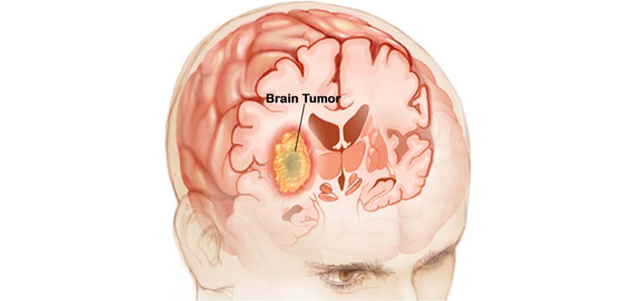
A brain tumour is an abnormal cell growth in the brain's tissues. Brain tumours can be benign (no cancer cells) or malignant (fast-growing cancer cells). Some of them are primary brain tumours, meaning they begin in the brain. Others are metastatic, which means they begin elsewhere in the body and spread to the brain.
As new cells replace old or damaged ones, normal cells proliferate in a regulated manner. Tumor cells multiply uncontrolled for reasons that are unknown.
A primary brain tumour is a benign tumour that begins in the brain and seldom spreads to other regions of the body. Primary brain tumours can be either benign or cancerous.
A benign brain tumour develops slowly, has well-defined borders, and spreads only infrequently. Benign tumours can be life threatening if they are placed in a key region, despite the fact that their cells are not cancerous.
A malignant brain tumour spreads to neighbouring brain regions, develops swiftly, and has irregular borders. Malignant brain tumours, despite their common name, do not meet the criteria of cancer since they do not spread to organs outside of the brain and spine.
Metastatic (secondary) brain tumours start out as cancer in another part of the body and then spread to the brain. When cancer cells are transported through the bloodstream, they develop tumours. Lung and breast cancers are the most prevalent malignancies that spread to the brain.
A brain tumour, whether benign, malignant, or metastatic, can all be life-threatening. The brain can't expand to make place for a growing mass since it's encased in a bony skull. The tumour compresses and displaces normal brain tissue as a result.
Some brain tumours cause the cerebrospinal fluid (CSF) that circulates around and through the brain to become clogged. This obstruction raises intracranial pressure and can cause the ventricles to expand (hydrocephalus). Swelling is a symptom of certain brain tumours (edema). The "mass effect" is caused by the size, pressure, and swelling of the body, which causes many of the symptoms.
Disease Signs and Symptoms:
Tumors can cause damage to the brain by killing healthy tissue, squeezing healthy tissue, or raising intracranial pressure. The kind, size, and location of the tumour in the brain all influence the symptoms. Symptoms in general include:
- Seizures with headaches that seem to get worse in the morning
- stumbling, dizziness, and walking difficulties
- issues with speech (e.g., difficulty finding the right word)
- irregular eye movements, visual difficulties
- Increased intracranial pressure due to weakness on one side of the body produces sleepiness, headaches, nausea and vomiting, and slow reactions.
The following are examples of specific symptoms:
Behavioral and emotional problems; poor judgement, motivation, or inhibition; decreased sense of smell or visual loss; paralysis on one side of the body; lower mental ability and memory loss are all possible adverse effects of frontal lobe tumours.
Parietal lobe tumours can cause difficulty with speaking, writing, drawing, and naming, as well as lack of recognition, spatial impairments, and eye-hand coordination.
Vision loss in one or both eyes, visual field cuts, fuzzy vision, illusions, and hallucinations are all possible symptoms of occipital lobe tumours.
Temporal lobe tumours can cause issues with speaking and interpreting language, as well as short- and long-term memory.
aggressiveness on the rise
Behavioral and emotional problems, trouble speaking and eating, tiredness, hearing loss, muscular weakness on one side of the face (e.g., head tilt, crooked grin), uncoordinated walking, drooping eyelid or double vision, and vomiting are all symptoms of brainstem tumours.
Increased hormone secretion (Cushing's Disease, acromegaly), cessation of menstruation, irregular milk secretion, and diminished libido are all possible side effects of pituitary gland tumours.
Disease Causes:
hereditary illnesses, such as neurofibromatosis, extended exposure to pesticides, industrial solvents, and other toxins cancer elsewhere in the body
ZapX brain surgery
ZAP-X Gyroscopic Radiosurgery: ZAP-X Gyroscopic Radiosurgery or ZapX brain surgery is a state-of-the-art technique designed to accurately identify and manage brain tumors. ZAP-X employs a novel self-shielded, gyroscopic linear accelerator architecture in contrast to more traditional techniques. As a result, it may target tumors from every direction surrounding the head, increasing its effectiveness and precision.
ZAP-X has a high success rate and little long-term negative effects, making it a viable treatment for a variety of illnesses. Because of ZAP-X's accuracy, specific treatments for a range of brain-related disorders are available.
Country wise cost comparison for Brain Tumor Surgery:
| Country | Cost |
|---|---|
| India | $6030 |
| Thailand | $25618 |
| United Arab Emirates | $30586 |
| Canada | $103666 |
Treatment and Cost
30
Total Days
In Country
- 5 Day in Hospital
- 2 No. Travelers
- 25 Days Outside Hospital
Treatment cost starts from
$0
Popular Hospital & Clinic
Featured Hospital
0 Hospitals
Related Packages
More Related Information
Some of the top rated hospitals are:
- Turkey
- Kolan International Hospital, Sisli
- Istinye University Bahcesehir Liv Hospital
- Istinye University Medical Park Gaziosmanpasa Hospital
- I.A.U VM Medical Park Florya Hospital
- Altinbas University Medical Park Bahcelievler Hospital
- Medical Park Antalya Hospital
- Medical Park Tarsus Hospital, Mersin
- Thailand
- Bangpakok 9 International Hospital
- Bumrungrad International Hospital
- Bangkok Hospital
- Bangkok International Hospital
- Samitivej Hospital
- BNH Hospital
- Aek Udon International Hospital
- Phuket International Hospital
- Bangkok Christian Hospital
- Thonburi Hospital
- Kasemrad Hospital Sriburin
- Canada
- Toronto General Hospital
- Jewish General Hospital
- Montreal General Hospital (McGill University Health Centre)
- Royal Jubilee Hospital (RJH)
- The Royal Victoria Hospital (McGill University Health Centre)
- Centre hospitalier de l’Université de Montréal (CHUM)
- Victoria General Hospital
- St Michaels Hospital Toronto
- Hamilton General Hospital
- MCMASTER UNIVERSITY MEDICAL CENTRE
- University of Ottawa Heart Institute
- Italy
Frequently Asked Questions
In the United Arab Emirates, brain tumor treatment typically costs $25,000. In the United Arab Emirates, there are numerous JCI and TEMOS approved hospitals that provide treatment for brain tumors.
The cost of treating brain tumors in the United Arab Emirates varies depending on the facility. A complete package that includes all costs associated with the patient's care and investigations is provided by some of the top hospitals for brain tumor treatment. The costs associated with hospitalization, surgery, nursing care, medications, and anesthesia are typically included in the treatment cost. The cost of brain tumour treatment in the United Arab Emirates can be raised by a number of factors, such as an extended hospital stay and post-procedural problems.
The price of brain tumor treatment in Dubai is contingent upon several aspects. The price of medications, anesthesia, hospital stays, and the surgeon's fee are all included in the cost of treatment in the kingdom. Longer hospital stays and problems following surgery are two factors that could drive up the total cost of brain cancer treatment in the nation.
Other elements influencing the price of brain tumor surgery in Dubai include:
therapy type: The cost of each type of therapy varies based on the intricacy of the process used.
The length of hospital stay: The total number of days the patient spends in the hospital from the time of admission until their discharge determines how much their brain tumor treatment will cost.
Patient status following surgery: If a patient experiences any complications following surgery, more care will be given to help stabilize their condition. To prevent complications, the underlying condition may occasionally need to be addressed before the surgery is carried out. This extra care raises the overall expense of the procedure.
Conditioning required before treatment: If additional therapy is needed prior to surgery, the cost of the procedure may increase.
Expertise of the specialists: A significant portion of the overall cost of the surgery is attributed to the fees charged by a brain tumor surgeon. The total experience, credentials, success rate, and hospital affiliation of a surgeon all play a role in determining their cost.
Underlying medical conditions: Prior to surgery, additional testing and medication may be required to treat or control any underlying medical conditions.
Post-operative testing and monitoring: Following the procedure, a team of committed medical professionals will keep a close eye on the patient's condition and, if necessary, request tests.
Patient age: Because elderly patients may require longer recovery times, their care may be more expensive.
In the United Arab Emirates, there are a number of top hospitals for treating brain tumors. The following hospitals are among the top ones in the UAE for treating brain tumors:
NMC Royal Hospital
NMC Speciality Hospital, Al Nahda
American Hospital
Medeor 24X7 Hospital
Zulekha Hospital
Canadian Specialist Hospital
In addition to the cost of Brain Tumor Treatment, the patient can be required to pay a few additional daily fees. These fees cover daily meals and lodging away from the hospital. The additional fees may begin at USD $50 per person.
The following are some of the top cities in the United Arab Emirates for brain tumor treatment:
Abu Dhabi
Dubai
Sharjah
Some of the best medical professionals in the United Arab Emirates providing treatment for brain tumors include the following:
Dr. Mehandi Hassan Ansari
Dr. Ajit Kumar
Dr. Shankar Ayyappan Kutty
Dr. Rahul Amunje Mally
Dr. Arif Khan


